|

|
Laboratory Overview
Our laboratory focuses on advancing optical and histological methodologies for 3D characterization of tissue microarchitecture, vasculature, and innervation. By applying optical clearing methods, we render tissues—such as mouse and human intestine, pancreas, liver, and kidney—transparent, enabling detailed 3D visualization of fluorescently labeled structures. A key aspect of our research is integrating modern 3D histology (fluorescence and transmitted light imaging) with classic techniques, such as H&E and IHC staining, to enable multiplexed analysis of tissues in both health and disease.
==================================
New in 2023: Antifade technology for 3D & super-resolution imaging in high-n polymer
==================================
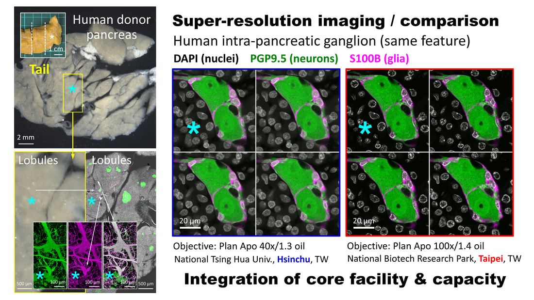
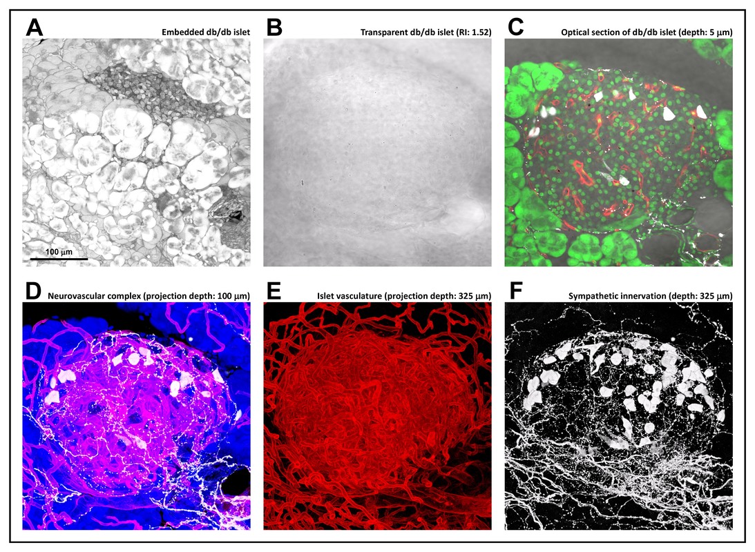
Our team recently developed a novel antifade method for 3D and super-resolution imaging, compatible with 3D Airyscan, STED, Lattice SIM, and NSPARC. This method uses solid high-refractive-index (high-n) polymers as an alternative to conventional immersion-based tissue clearing techniques.
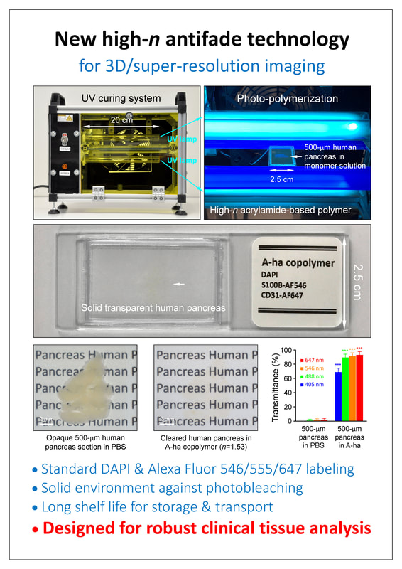
Rather than using high-n liquids, we embed fluorescently labeled tissues in a solid, acrylamide-based high-n polymer through photo-polymerization. Much like tissue solidification with paraffin for H&E histology or resin for electron microscopy, this solvent-free environment is optimized for clinical tissue analysis.
By limiting oxygen diffusion into the specimen, it significantly reduces photobleaching and minimizes signal loss during imaging, while providing extended shelf life for long-term storage, transport, and repeated fluorescence imaging.
==================================
Keywords: 3D histology; human pancreas early change/lesion; human pancreas & liver 3D neurohistology; high-n polymer, solid-state tissue clearing; antifade 3D & super-resolution imaging; 1080p HD video; 3D Airyscan; STED; Lattice SIM; NSPARC; 3D super-resolution islet imaging
==================================
Our laboratory focuses on advancing optical and histological methodologies for 3D characterization of tissue microarchitecture, vasculature, and innervation. By applying optical clearing methods, we render tissues—such as mouse and human intestine, pancreas, liver, and kidney—transparent, enabling detailed 3D visualization of fluorescently labeled structures. A key aspect of our research is integrating modern 3D histology (fluorescence and transmitted light imaging) with classic techniques, such as H&E and IHC staining, to enable multiplexed analysis of tissues in both health and disease.
==================================
New in 2023: Antifade technology for 3D & super-resolution imaging in high-n polymer
==================================
Our team recently developed a novel antifade method for 3D and super-resolution imaging, compatible with 3D Airyscan, STED, Lattice SIM, and NSPARC. This method uses solid high-refractive-index (high-n) polymers as an alternative to conventional immersion-based tissue clearing techniques.
Rather than using high-n liquids, we embed fluorescently labeled tissues in a solid, acrylamide-based high-n polymer through photo-polymerization. Much like tissue solidification with paraffin for H&E histology or resin for electron microscopy, this solvent-free environment is optimized for clinical tissue analysis.
By limiting oxygen diffusion into the specimen, it significantly reduces photobleaching and minimizes signal loss during imaging, while providing extended shelf life for long-term storage, transport, and repeated fluorescence imaging.
==================================
Keywords: 3D histology; human pancreas early change/lesion; human pancreas & liver 3D neurohistology; high-n polymer, solid-state tissue clearing; antifade 3D & super-resolution imaging; 1080p HD video; 3D Airyscan; STED; Lattice SIM; NSPARC; 3D super-resolution islet imaging
==================================
1080pHD video: 3D/Airyscan super-resolution imaging of human intrapancreatic ganglion in high-n polymer
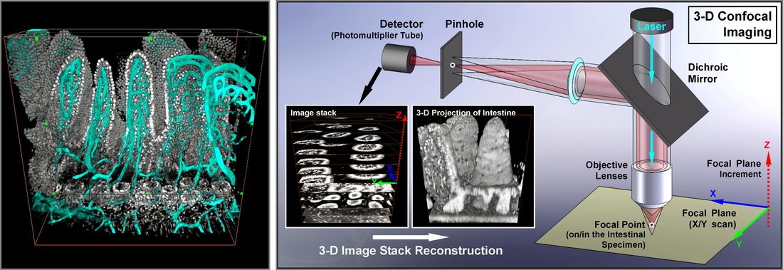
Toward a 3D new world of modern histology
3D images are essential for the study of tissue networks to characterize their morphologies in space. We establish tissue labeling and microscopic tools for high-resolution 3D imaging of mouse and human tissues.
Our featured cover images and illustrations
Integration of classic and in-depth tissue imaging (demonstration -- human pancreatic duct lesion)
Team and collaborators
                                PI: Dr. Shiue-Cheng (Tony) Tang; Dr. Pankaj J. Pasricha; Dr. Yuan-Chiang Chung; Dr. Chia-Ning Shen; Dr. Michael German; Dr. Chester Chamberlain; Dr. Luc Baeyens; David Scheel; Dr. Yu-Wen Tien; Dr. Yung-Ming Jeng; Dr. Chih-Yuan Lee; Dr. Shien-Tung Pan; Dr. Ming-Yin Shen; Dr. Yung-Chi Hou, Dr. Tsung-Lin Yang; Dr. Jyuhn-Huarng Juang; Dr. Ya-Yuan Fu; Dr. Tzu-En Hua; Dr. Pei-Yu Lin; M.Sc. Yu-Chen Chiu; Dr. Yuan-An Liu; M.Sc. Shih-Jung Peng; M.Sc. Chia-Tung Hsu; Dr. Chien-Chang Huang; Dr. Hung-Jen Chien; M.Sc. Ya-Hsien Chou; Dr. Fu-Ting Hsiao; M.Sc. Mei-Hsin Chung; M.Sc. Tzu-Hui Huang; Dr. Li-Wen Lo; Dr. Chien-Chia Chen
|
At the Movies: 3-D Technology & Gastrointestinal Histology |
|
|
|
|
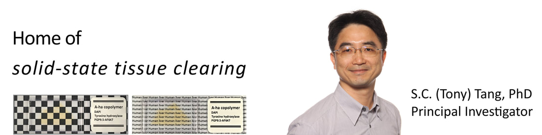
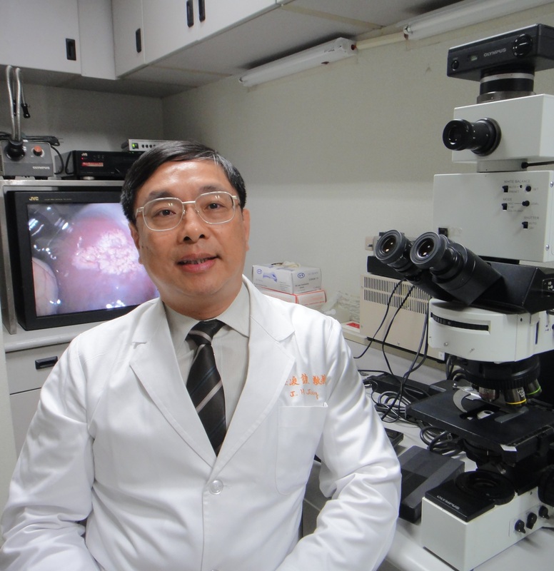
About the PI
Shiue-Cheng (Tony) Tang, Ph.D.
sctang@life.nthu.edu.tw
Department of Medical Science
Institute of Biotechnology
National Tsing Hua University
101, Sec. 2, Kuang Fu Rd., Hsinchu, Taiwan, 30013
Tel: 886-3-574-2465
2017 American Gastroenterological Association (AGA) Fellow
2015 Visiting Scholar, Diabetes Center, UCSF
2012-present
Professor, National Tsing Hua University, Institute of Biotechnology
Professor, National Tsing Hua University, Department of Medical Science
Professor, National Tsing Hua University, Department of Chemical Eng (joint appointment)
2008-2011
Associate Professor, National Tsing Hua University, Taiwan, Dept. of Chemical Eng
2005-2008
Assistant Professor, National Tsing Hua University, Taiwan, Dept. of Chemical Eng
2005 Jan-Jul
Assistant Professor, Nanyang Technological University, Singapore, Div. of Chemical & Biomolecular Engineering, School of Chemical & Biomedical Engineering
2004
Postdoctoral Training, Pediatric Gastroenterology, Stanford University School of Medicine
1998-2003
PhD Training, Chemical and Biomolecular Engineering, Georgia Institute of Technology
Our publications on 3-D histology
|
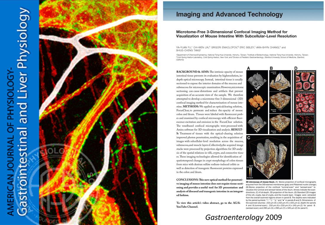
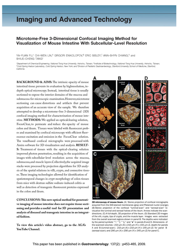
1. Fu YY, Lin CW, Enikolopov G, Sibley E, Chiang AS, and Tang SC. Microtome-free 3-dimensional confocal imaging method for visualization of mouse intestine with subcellular-level resolution. Gastroenterology. 137(2): p453-465, 2009.
|
|
|
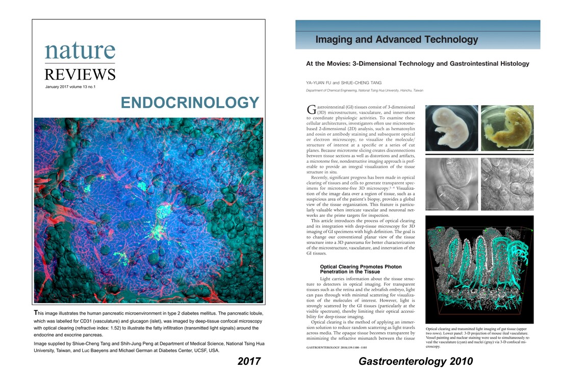
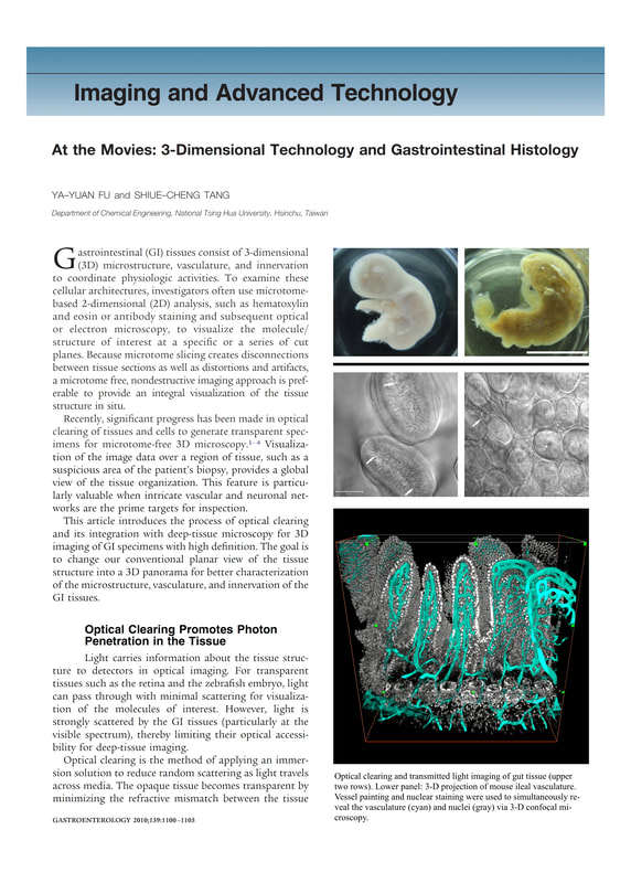
2. Fu YY and Tang SC. At the movies: 3-dimensional technology and gastrointestinal histology. Gastroenterology, 139(4): p1100-1105, 2010.
|
|
|
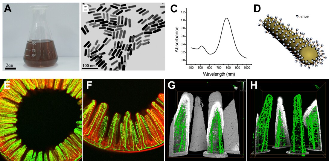
3. Tang SC, Fu YY, Lo WF, Hua TE, and Tuan HY. Vascular labeling of luminescent gold nanorods enables 3-D microscopy of mouse intestinal capillaries. ACS Nano, 4(10):6278-84, 2010.
|
|
|
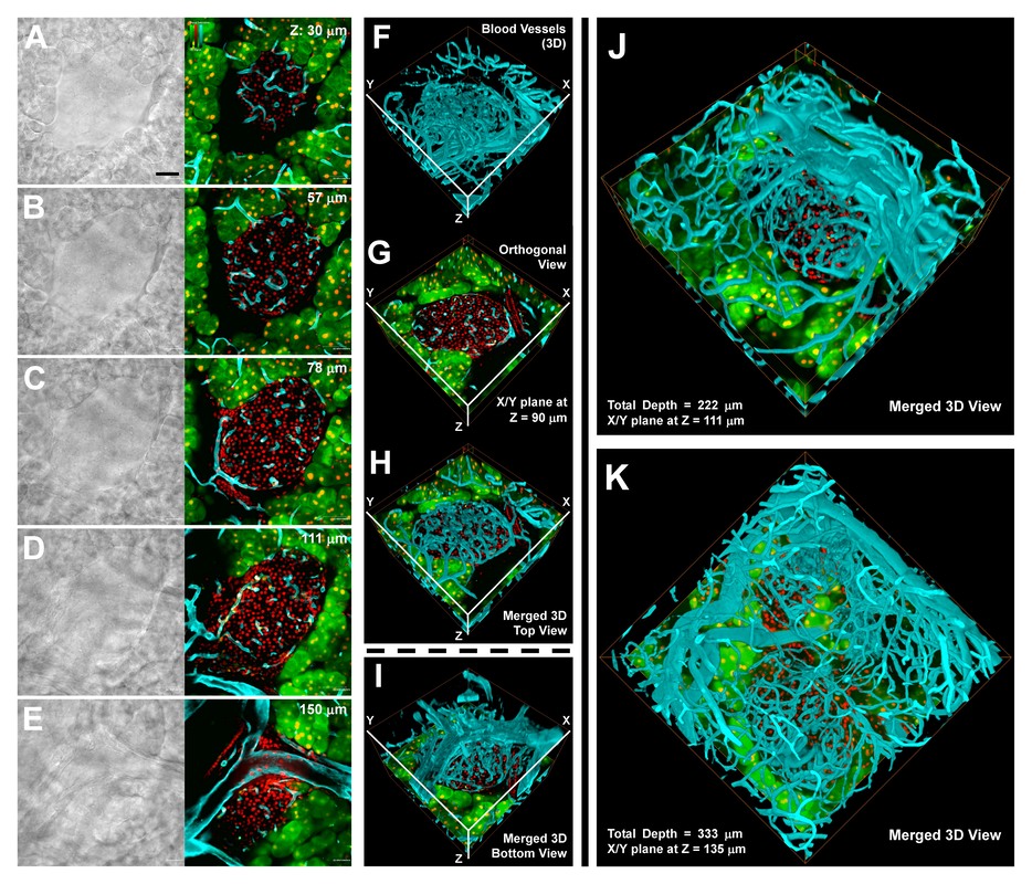
4. Fu YY and Tang SC. Optical clearing facilitates integrated 3D visualization of mouse ileal microstructure and vascular network with high definition. Microvascular Research, 80(3):512-21, 2010.
|
|
|
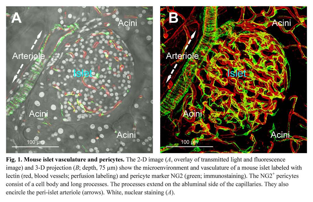
5. Fu YY, Lu CH, Lin CW, Juang JH, Enikolopov G, Sibley E, Chiang AS, Tang SC. Three-dimensional optical method for integrated visualization of mouse islet microstructure and vascular network with subcellular-level resolution. Journal of Biomedical Optics. 15(4), Article Number: 046018, 2010.
|
|
|
6. Tseng SJ, Lee YH, Chen ZH, Lin HH, Lin CY, and Tang SC. Integration of optical clearing and optical sectioning microscopy for three dimensional imaging of natural biomaterial scaffolds in thin sections. Journal of Biomedical Optics. 14(4), Article Number: 044004, 2009.
|
|
|
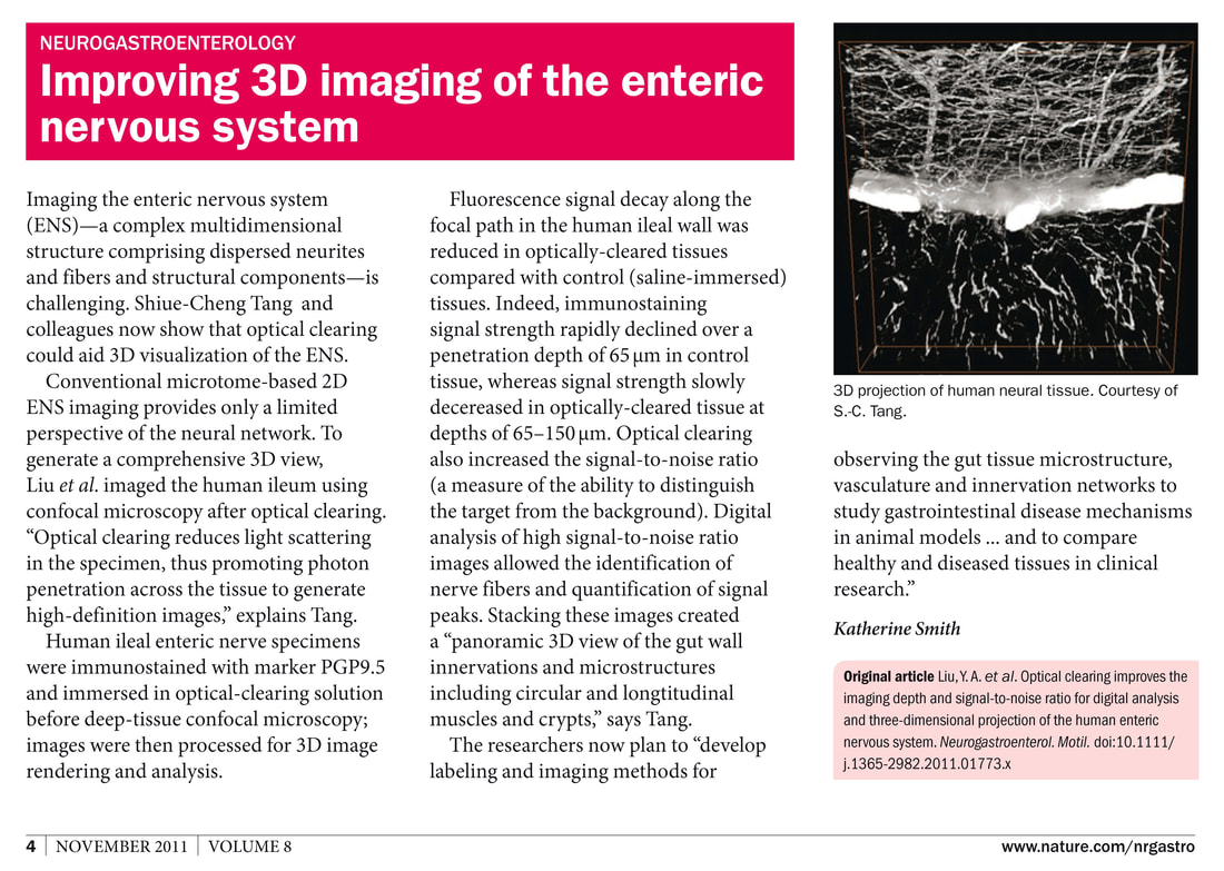
7. Liu YA, Chen Y, Chiang AS, Peng JS, Pasricha PJ, and Tang SC. Optical clearing improves the imaging depth and signal-to-noise ratio for digital analysis and 3-dimensional projection of the human enteric nervous system. Neurogastroenterology & Motility. 23:e446-457, 2011.
|
|
Highlighted by Nature Reviews Gastroenterology & Hepatology 8: 600; “Improving 3D imaging of the enteric nervous system” https://www.nature.com/articles/nrgastro.2011.167
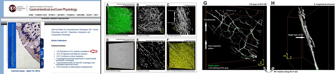
8. Liu YA, Chung YC, Pan ST, Hou YC, Peng SJ, Pasricha PJ, and Tang SC. 3-D illustration of network orientations of interstitial cells of Cajal subgroups in human colon as revealed by deep-tissue imaging with optical clearing. American Journal of Physiology - Gastrointestinal and Liver Physiology. 302:G1099-G1110, 2012. Selected as featured article: "3-D illustration of ICC network orientations"
|
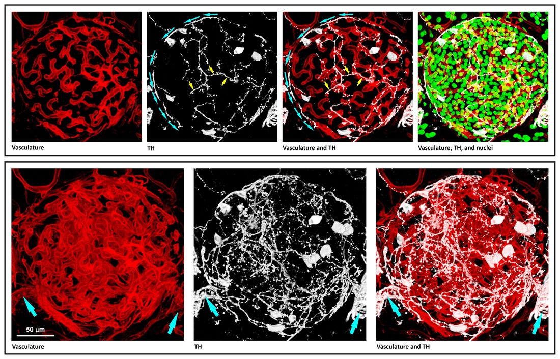
9. Chiu YC, Hua TE, Fu YY, Pasricha PJ, and Tang SC. 3-D imaging and illustration of the perfusive mouse islet sympathetic innervation and its remodelling in injury. Diabetologia. 55:3252-3261, 2012. Featured article.
|
|
Accompanied by a commentary: “Islet nerves in focus – defining their neurobiological and clinical role” https://pubmed.ncbi.nlm.nih.gov/23001378
|
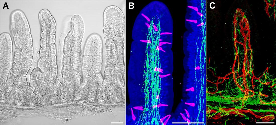
10. Liu YA, Chung YC, Shen MY, Kuo CW, Li WL, Pasricha PJ, Tang SC. Whole-villus neurohistology with optical clearing. Gastroenterology, 146 (5), Suppl. 1, May 2014, Pages S-120. DDW abstract publication.
|
|
|
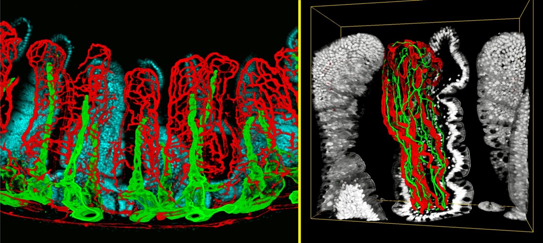
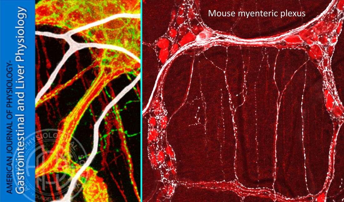
11. Fu YY, Peng SJ, Lin HY, Pasricha PJ, and Tang SC. 3-D imaging and illustration of mouse intestinal neurovascular complex. American Journal of Physiology - Gastrointestinal and Liver Physiology. 304:G1-G11, 2013. Cover image.
|
|
|
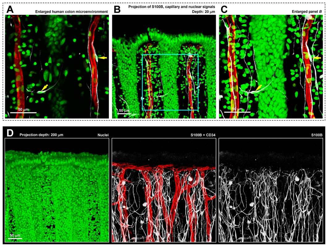
12. Liu YA, Chung YC, Pan ST, Shen MY, Hou YC, Peng SJ, Pasricha PJ, and Tang SC. 3-D imaging, illustration, and quantitation of enteric glial network in transparent human colon mucosa. Neurogastroenterology & Motility. 25:e324-338, 2013.
|
|
|
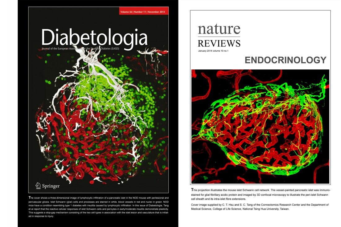
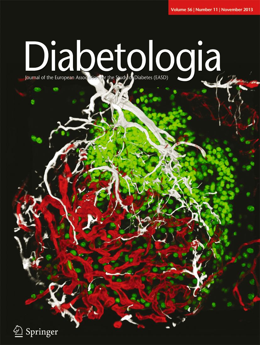
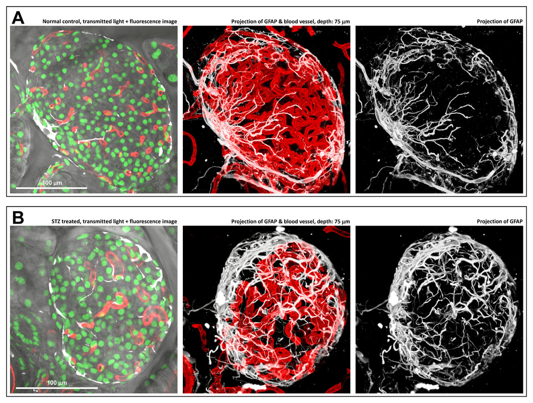
13. Tang SC, Chiu YC, Hsu CT, Peng SJ, and Fu YY. Plasticity of Schwann cells and pericytes in response to islet injury in mice. Diabetologia. 56:2424-2434, 2013. Cover image of November issue.
|
|
|
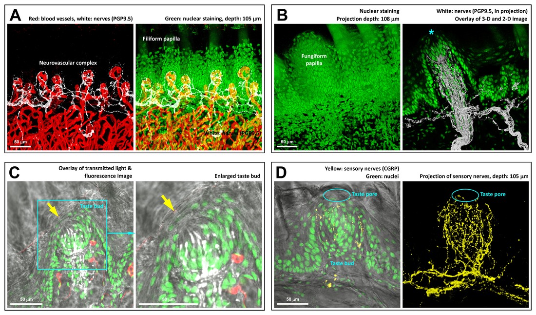
14. Hua TE, Yang TL, Yang WC, Liu KJ, and Tang SC. 3-D neurohistology of transparent tongue in health and injury with optical clearing. Frontiers in Neuroanatomy, 7:36, 2013.
|
|
|
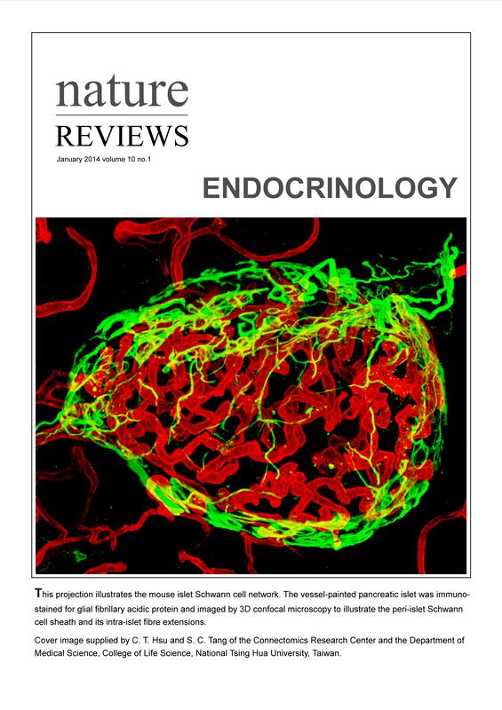
15. Hsu CT and Tang SC. Nature Reviews Endocrinology, Cover image 2014 Jan-Dec: Mouse islet Schwann cell network. Winner of 2014 Nature Reviews cover image competition. https://www.nature.com/nrendo/volumes/10/issues/1
|
|
|
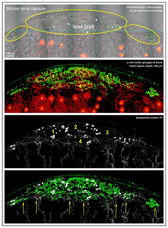
16. Juang JH, Peng SJ, Kuo CH, and Tang SC. 3D islet graft histology: panoramic imaging of neural plasticity in sympathetic reinnervation of transplanted islets under the kidney capsule. American Journal of Physiology - Endocrinology and Metabolism, 306:E559-E570, 2014.
|
|
|
17. Tang SC, Peng SJ, and Chien HJ. Imaging of the islet neural network. Diabetes, Obesity and Metabolism, 16 (Suppl. 1):77-86. 2014. Review.
|
|
|
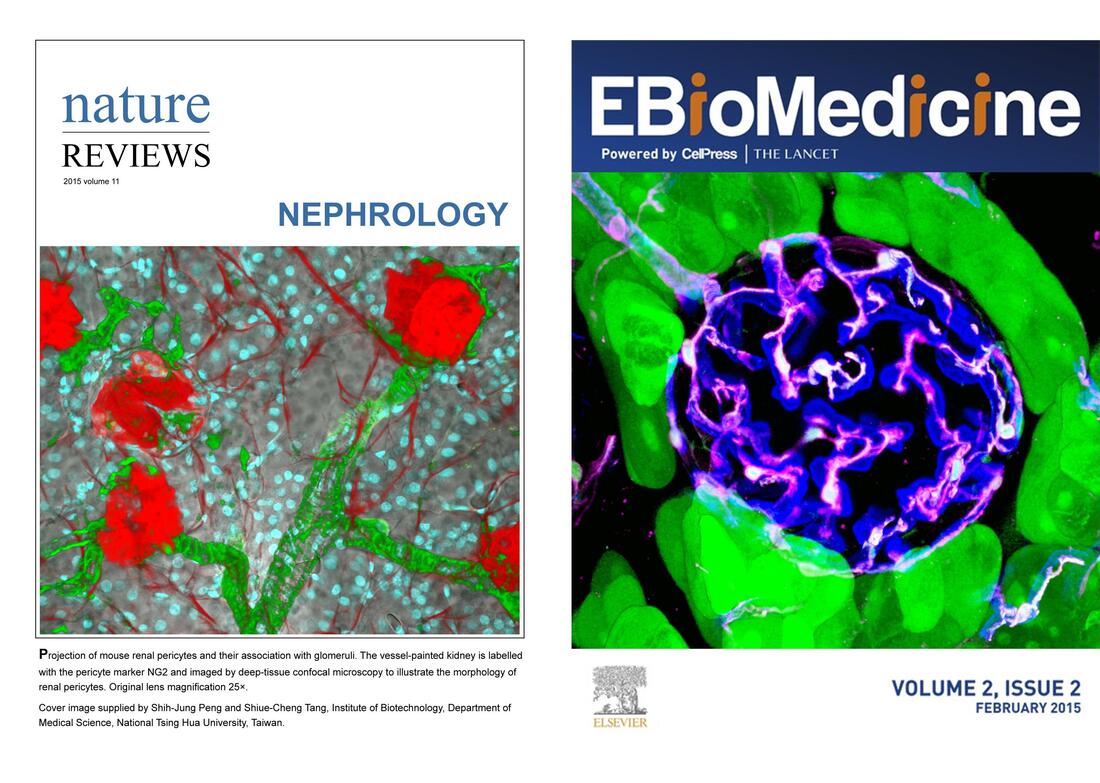
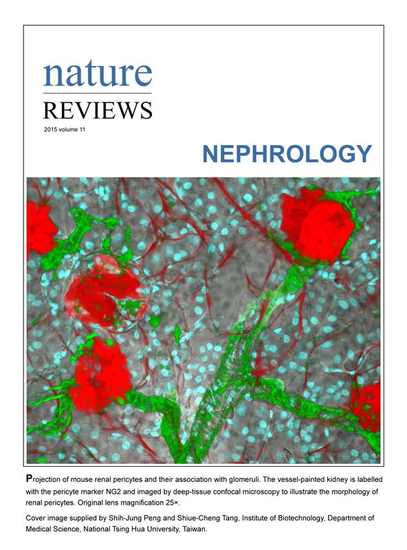
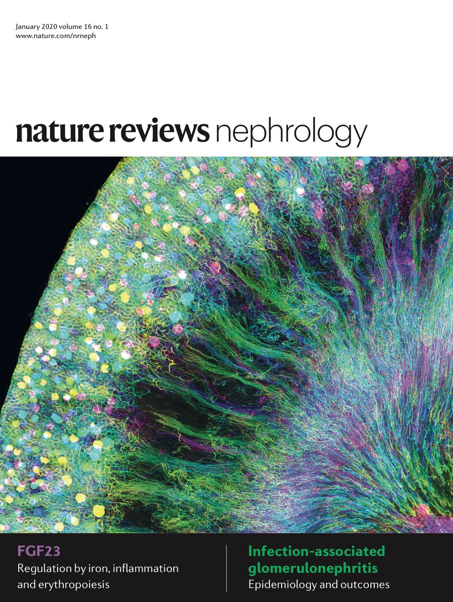
18. Peng SJ and Tang SC*. Nature Reviews Nephrology, Cover image 2015 Jan-Dec, all twelve issues: Projection of mouse renal pericytes and their association with glomeruli. https://www.nature.com/nrneph/volumes/11/issues/12
|
|
|
19. Liu YA, Chung YC, Shen MY, Pan ST, Kuo CW, Peng SJ, Pasricha PJ, and Tang SC. Perivascular interstitial cells of Cajal in human colon. Cellular and Molecular Gastroenterology and Hepatology. American Gastroenterological Association (AGA) journal, 1:102-119, 2015.
|
|
|
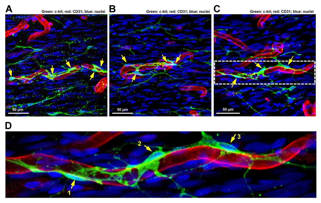
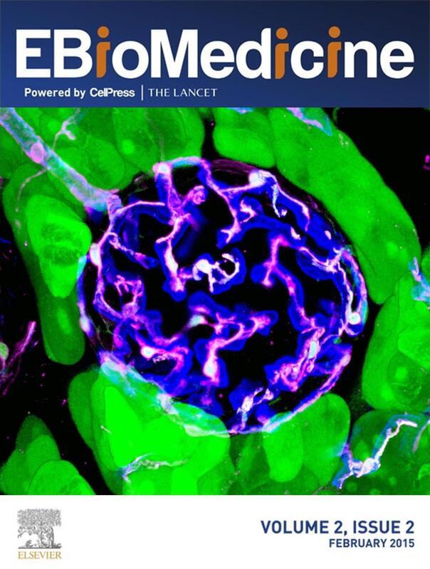
20. Juang JH, Kuo CH, Peng SJ, and Tang SC. 3-D imaging reveals participation of donor islet Schwann cells and pericytes in islet transplantation and graft neurovascular regeneration. EBioMedicine, 2:109-119, 2015. Cover image of February issue.
|
|
|
21. Chien HJ, Peng SJ, Hua TE, Kuo CH, Juang JH, and Tang SC. 3-D imaging of islets in obesity: formation of the islet-duct complex and neurovascular remodeling in young hyperphagic mice. International Journal of Obesity, 40: 685-697, 2016.
|
|
|
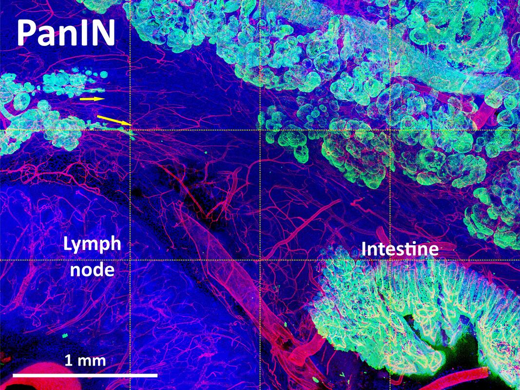
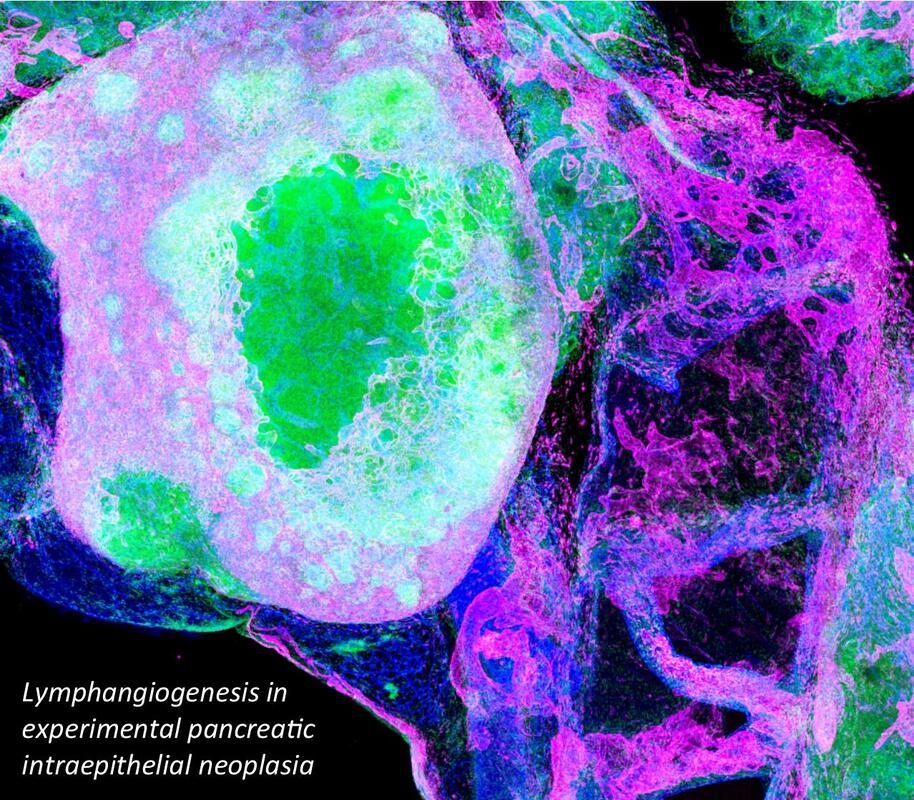
22. Lin PY, Peng SJ, Shen CN*, Pasricha PJ, and Tang SC*. PanIN-associated pericyte, glial, and islet remodeling in mice revealed by 3-D pancreatic duct lesion histology. American Journal of Physiology - Gastrointestinal and Liver Physiology, 311: G412–G422, 2016.
|
|
|
Nestin-GFP mouse showing the nestin-positive cell network in the small intestine. The nestin-GFP (green) cells form a network in the longitudinal muscle-myenteric plexus layer, submucosal layer, and also inside the villi. Some nestin-GFP–expressing cells are present around the vasculature, visualized using DiD vessel painting technique (cyan).
|
23. Kulkarni S, Micci MA, Leser J, Shin C, Tang SC, Fu YY, Liu L, Li Q, Saha M, Li C, Enikolopov G, Becker L, Rakhilin N, Anderson M, Shen X, Dong X, Butte MJ, Song H, Southard-Smith EM, Kapur RP, Bogunovic M, Pasricha PJ. Adult enteric nervous system in health is maintained by a dynamic balance between neuronal apoptosis and neurogenesis. Proc Natl Acad Sci USA,114: E3709-E3718, 2017. (Contribution: application of 3-D histology to investigate enteric nervous system)
|
|
24. Tang SC*, Peng SJ, Baeyens L, and German MS. Nature Reviews Endocrinology, Cover image 2017 Jan-Dec: Human pancreatic microenvironment in type 2 diabetes mellitus. Winner of 2017 Nature Reviews cover image competition. www.nature.com/nrendo/volumes/13/issues/1
|

|
|
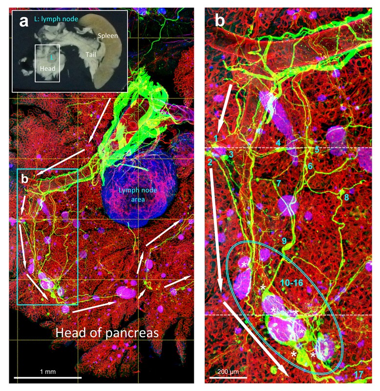
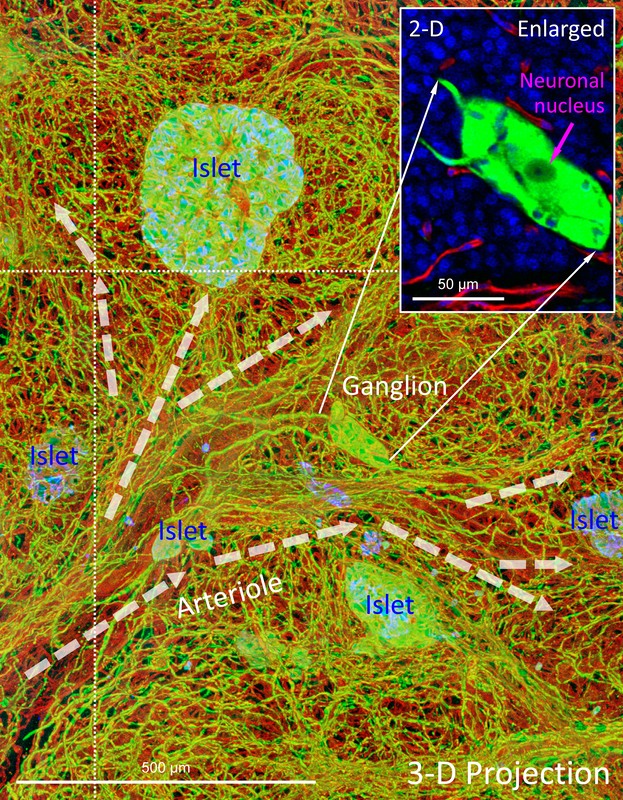
25. Tang SC*, Shen CN*, Lin PY, Peng SJ, Chien HJ, Chou YH, Chamberlain CE, and Pasricha PJ. Pancreatic neuro-insular network in young mice revealed by 3-D panoramic histology. Diabetologia. 61:158-167, 2018. Back-to-back featured article.
|
|
|
26. Tang SC*, Baeyens L, Shen CN, Peng SJ, Chien HJ, Scheel DW, Chamberlain CE, and German MS*. Human pancreatic neuro-insular network in health and fatty infiltration. Diabetologia. 61:168-181, 2018. Back-to-back featured article.
|
|
|
27. Tang SC*, Jessup CF*, and Campbell-Thompson M*. The role of accessory cells in islet homeostasis. Current Diabetes Reports. 18(11):117, 2018. Review.
|
|
|
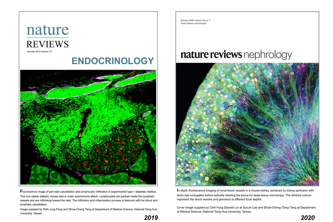
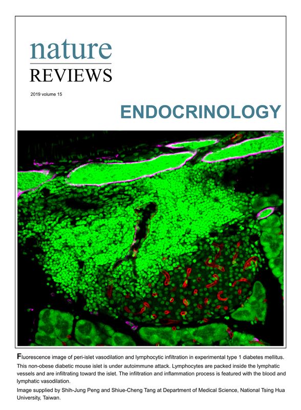
28. Peng SJ and Tang SC*. Nature Reviews Endocrinology, Cover image 2019 Jan-Dec: Peri-islet vasodilation and lymphocytic infiltration in experimental type 1 diabetes mellitus. Winner of 2019 Nature Reviews cover image competition. www.nature.com/nrendo/volumes/15/issues/1
|
|
|
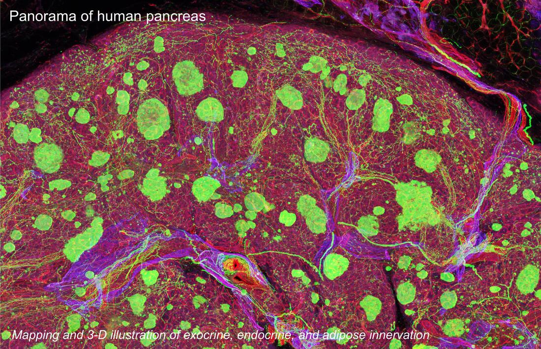
29. Chien HJ, Chiang TC, Peng SJ, Chung MH, Chou YH, Lee CY, Jeng YM, Tien YW, and Tang SC*. Human pancreatic afferent and efferent nerves: mapping and 3-D illustration of exocrine, endocrine, and adipose innervation. American Journal of Physiology - Gastrointestinal and Liver Physiology, 317: G694–G706, 2019.
|
|
|
30. Shen CN, Goh KS, Huang CR, Chiang TC, Lee CY, Jeng YM, Peng SJ, Chien HJ, Chung MH, Chou YH, Hsieh CC, Kulkarni S, Pasricha PJ, Tien YW*, and Tang SC*. Lymphatic vessel remodeling and invasion in pancreatic cancer progression. EBioMedicine, 47:98-113, 2019.
|
|
|
31. Lin CY and Tang SC*. Nature Reviews Nephrology, Cover image 2020 Jan-Dec, all twelve issues: In-depth fluorescence imaging of renal blood vessels. Winner of 2020 Nature Reviews cover image competition. www.nature.com/collections/fifigibgci ; www.nature.com/nrneph/volumes/16/issues/1
|
|
|
32. Campbell-Thompson M and Tang SC. Pancreas optical clearing and 3-D microscopy in health and diabetes. Frontiers in Endocrinology, 12: Article 644826, 2021. Review.
|
|
|
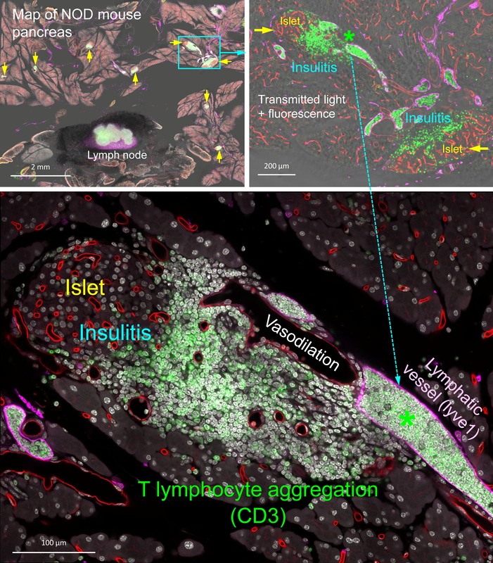
|
In-depth imaging of optically cleared NOD mouse pancreas with insulitis. Overlay of transmitted light and fluorescence signals identifies the Lyve1+ lymph node (filled with CD3+ T lymphocytes and surrounded by fats, image at the top) and locations of insulitic islets. Islets with insulitis are shown with blood vessels (red), lymphatic vessels (magenta), and nuclei (white). CD3+ T lymphocytes are identified around the islets and congregated in the lymphatic vessels.
|
|
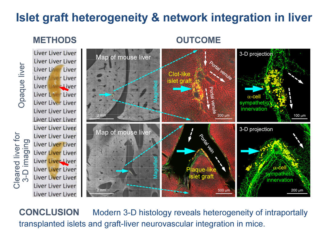
33. Chen CC, Peng SJ, Wu PY, Chien HJ, Lee CY, Chung MH, and Tang SC. Heterogeneity and neurovascular integration of intraportally transplanted islets revealed by 3-D mouse liver histology. American Journal of Physiology - Endocrinology and Metabolism, 320:E1007-E1019, 2021.
|
|
|
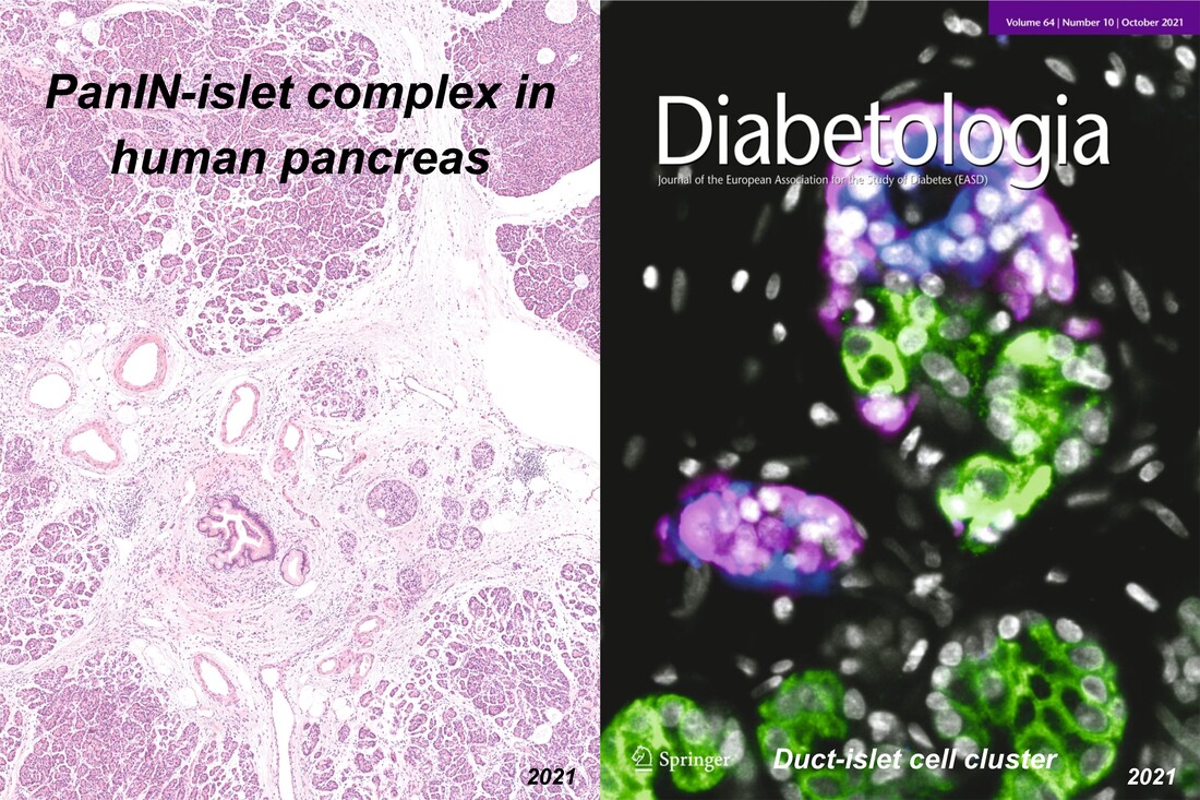
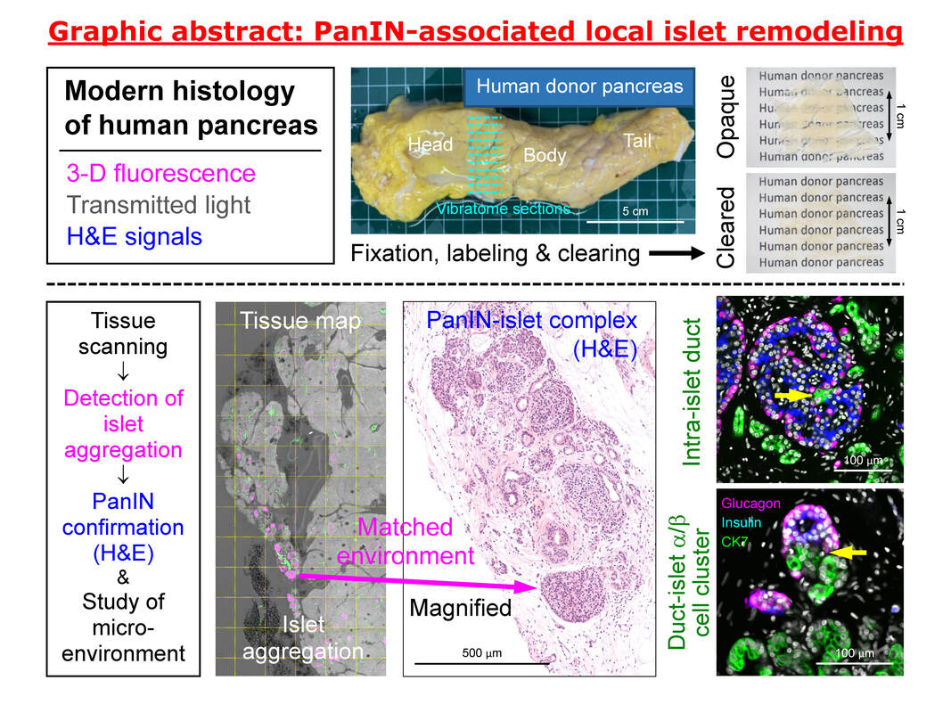
34. Tien YW, Chien HJ, Chiang TC, Chung MH, Lee CY, Peng SJ, Chen CC, Chou YH, Hsiao FT, Jeng YM, and Tang SC. Local islet remodeling associated with duct lesion-islet complex in adult human pancreas. Diabetologia, 64:2266-2278, 2021. Cover image.
|
|
35. Shiue-Cheng Tang. 2021 US Patent 11,009,433: Composition and method for solid-state tissue clearing. https://uspto.report/patent/grant/11,009,433
|
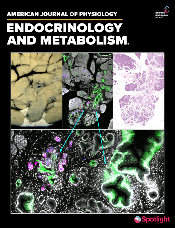
36. Chung MH, Chien HJ, Peng SJ, Chou YH, Chiang TC, Chang HP, Lee CY, Chen CC, Jeng YM, Tien YW*, Tang SC*. Multimodal 3-D/2-D human islet and duct imaging in exocrine and endocrine lesion environment: associated pancreas tissue remodeling. American Journal of Physiology - Endocrinology and Metabolism, 323:E354-E365, 2022.
|
|
|
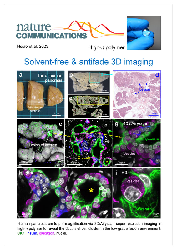
37. Hsiao FT, Chien HJ, Chou YH, Peng SJ, Huang TH, Lo LW, Sheng CN, Chang HP, Lee CY, Chen CC, Jeng YM, Tien YW, Tang SC*. Transparent tissue in solid state for solvent-free and antifade 3D imaging. Nature Communications, 14:3395, 2023. https://www.nature.com/articles/s41467-023-39082-4
|
|
|
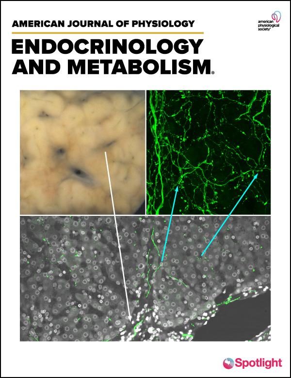
38. Chen CC, Peng SJ, Chou YH, Lee CY, Lee PH, Hu RH, Ho MC, Chung MH, Hsiao FT, Tien YW, Tang SC*. Human liver afferent and efferent nerves revealed by 3-D/Airyscan super-resolution imaging. American Journal of Physiology - Endocrinology & Metabolism, 326:E107-E123, 2024. https://journals.physiology.org/doi/abs/10.1152/ajpendo.00205.2023
|
|

|
|
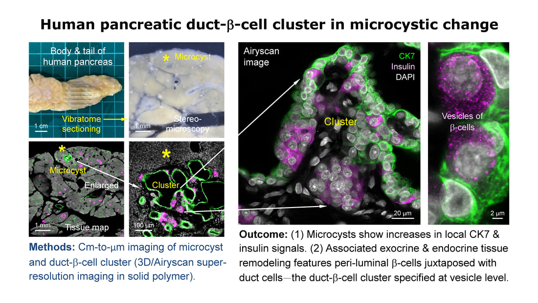
39. Lee CY, Kuo TC, Chou YH, Peng SJ, Hsiao FT, Chung MH, Lo LW, Shen CN, Chien HJ, Chang HP, Chen CC, Jeng YM, Tien YW*, Tang SC*. 3D imaging resolves human pancreatic duct-beta-cell clusters during cystic change. Diabetes, 74:734-748, 2025. https://pubmed.ncbi.nlm.nih.gov/39787388/
Accompanied by a commentary: “Bend It Like Occam: Ductal Origin of New Islet Cells in Human Pancreas After Injury.” Diabetes, 74:682-684, 2025. https://pubmed.ncbi.nlm.nih.gov/40258166/
Accompanied by a commentary: “Bend It Like Occam: Ductal Origin of New Islet Cells in Human Pancreas After Injury.” Diabetes, 74:682-684, 2025. https://pubmed.ncbi.nlm.nih.gov/40258166/